
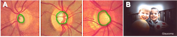
A) Figure showing a close up view of the optic nerve head in the back of the eye. Progressive enlargement of the optic nerve head “cup” (green outline) over time is indicative of damage to the nerve. (Figures from www.eyemdlink.com). B) A visual scene as might be viewed by a patient with glaucoma. (figure from www.nei.nih.gov)
In glaucoma, the retinal ganglion cells that eventually form the optic nerve (which in turn sends visual information from the retina to the brain) slowly die over time. At first, side (peripheral) vision is lost. If glaucoma is not treated, vision loss may continue, leading to total blindness over time. There are many types of glaucoma and the exact causes are still unknown but frequently high eye (intraocular) pressure is often associated with this condition. Current treatment is with medications and surgical techniques aimed at lowering intraocular pressure.