
Approaches to Restore Vision using a Visual Neuroprosthesis
A prosthesis can be described as a man-made device intended to replace the function of a damaged body part. To help visually impaired patients, a visual prosthesis is designed to electrically stimulate and replace the component of the normal visual pathway such as the retina of the eye, the optic nerve, or in the visual cortex of the brain.
Click this image for a larger version
Image © Boston Retinal Implant Project
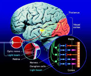
The visual system. Light is captured and focused onto the back surface of the eye (the retina; inset circle). Light detecting cells (rods and cones) along with other cell types (ganglion and bipolar cells) within the retina process visual information that in turn leaves the eye (via the optic nerve) to communicate with the visual cortex located in the back of the brain. Once in the cortex, visual information is further analyzed in order to generate visual perceptions that allow us to interpret what we see. Diagram courtesy of CPO Science, Peabody MA.
Click this image for a larger version
Image © Boston Retinal Implant Project
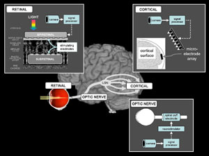
Summary diagram of the visual system and approaches to restore vision. In theory, any point along the visual pathway can be electrically stimulated and so represents a potential site at which a visual prosthesis could be implanted. The inset figures illustrate several approaches in detail. A) Schematic diagram of a retinal cross-section showing two methods of stimulating ganglion cells: epiretinal and subretinal (see section "description of the retinal approach" for more details). B) The optic nerve can be stimulated by implanting a cuff electrode around the nerve. The cuff electrode (located intracranially) and neurostimulator (placed beneath the skin) communicate wirelessly with an external processor and camera. C) The cortical approach incorporates a microelectrode array, which is placed either intracortically or on the cortical surface. By stimulating the visual cortex directly, this approach bypasses the retina and optic nerve.