
In vivo experiments (i.e. within a living organism) include the study of: 1) new surgical techniques to implant our retinal prosthesis, 2) the biocompatibility of the implanted device, and 3) the amount of electricity needed to stimulate retinal cells so that responses can be recorded from the visual cortex of the brain.
We have found that some materials tested in animals appear to be biocompatible and might be eventually used in a human implant. However, many materials used to cover the microelectrode stimulation array consistently become encapsulated by cells when implanted within the retina. This process of encapsulation represents a reaction of the body to the presence of a foreign object within biological tissues. It may potentially create a high resistance electrical barrier between the electrodes and the nerve cells we hope to stimulate. This in turn would require that the implanted device deliver more electricity, thus compromising its safety. This finding has led us to initiate other experiments designed to minimize the risk of encapsulation.
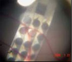
Magnified view of the back of the pig eye showing the surgical placement of an implanted stimulating array. Note the position of the array behind the retinal blood vessels.