
We are using new techniques to study the effects of blindness on brain development and function. Functional Magnetic Resonance Imagin (fMRI) allows the study of human brain activity in a non-invasive manner. Experiments using functional neuroimaging are aimed at studying visual cortex activity in response to different forms of blindness including recovery from stroke. Collaborative sites including the Center for Biomedical Imaging at Boston University Medical Center and MGH Martinos Center for Biomedical Imaging in Charlestown, MA.
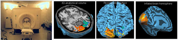
The General Electric 3 Tesla Scanner used for functional magnetic resonance imaging (fMRI). Images generated showing the activation within occipital visual cortex (axial slice projection), cortical surface reconstruction and inflated brain projection.