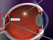
Since our the completion of our first phase of human clinical testing, we have developed a novel surgical approach in order to implant our retinal prosthesis in the eye. This represents an approach that is minimally invasive and a considerable improvement over more conventional surgical approaches being used today.

Description of surgical approach. Cross sectional view of the eye. Entry is made through two ports: one for light and a second for a retinal manipulating tool. A small pocket is made and an insertion flap is made by injecting fluid in order to separate the retina from the surface and create a potential space for the implant. In a second step, the implant is inserted through a corresponding site on the outside of the eye (referred to as an "ab externo" approach).
Surgical testing using animal models have demonstrated that we can implant our retina microchip into sub-retinal space safely and with reliable success using a combination of vitreoretinal surgery and ab externo procedure.
